Introduction to Gastroschisis
Gastroschisis is a rare congenital anomaly characterized by an abdominal wall defect, resulting in the intestine protruding outside the baby’s body. This condition requires immediate neonatal surgery to repair the defect and restore intestinal function.
Causes and Risk Factors
The exact cause of gastroschisis is unknown, but risk factors include young maternal age, low socioeconomic status, and exposure to environmental toxins during pregnancy, which may contribute to the development of this congenital anomaly.
Maternal Health and Fetal Development
Maternal health plays a crucial role in fetal development, and any factors that compromise maternal well-being may increase the risk of gastroschisis. Poor nutrition, inadequate prenatal care, and exposure to environmental toxins during pregnancy can all negatively impact fetal development.
Research suggests that maternal stress, anxiety, and depression may also contribute to an increased risk of gastroschisis. Furthermore, women who have a history of substance abuse or smoking during pregnancy are more likely to give birth to an infant with gastroschisis.
Fetal development is a complex process, and any disruptions to this process can result in congenital anomalies such as gastroschisis. It is essential for expectant mothers to prioritize their health and well-being during pregnancy to minimize the risk of complications and promote healthy fetal development.
Healthcare providers should work closely with expectant mothers to identify any potential risk factors and develop strategies to mitigate them, ultimately reducing the risk of gastroschisis and promoting optimal maternal and fetal health.
Genetic Predisposition
While the exact causes of gastroschisis are not yet fully understood, research suggests that genetic predisposition may play a role in the development of this condition. Studies have identified several genetic mutations that may contribute to an increased risk of gastroschisis.
Families with a history of gastroschisis or other abdominal wall defects are more likely to have an infant born with this condition. Additionally, certain genetic syndromes, such as omphalocele, have been linked to an increased risk of gastroschisis.
Further research is needed to fully understand the relationship between genetic predisposition and gastroschisis. However, it is clear that genetic factors can contribute to the development of this condition. Genetic counseling and testing may be beneficial for families with a history of gastroschisis or other abdominal wall defects.
Identifying genetic mutations associated with gastroschisis can help healthcare providers to develop targeted interventions and prevention strategies, ultimately reducing the incidence of this condition and improving outcomes for affected infants.
Prenatal Diagnosis and Management
Prenatal diagnosis of gastroschisis is crucial for optimal management. Advanced imaging techniques enable early detection, allowing for multidisciplinary care planning and preparation for neonatal surgery, thereby improving infant outcomes and reducing maternal anxiety.
Prenatal Ultrasound and Screening
Prenatal ultrasound is the primary diagnostic tool for detecting gastroschisis. A mid-pregnancy ultrasound scan can identify the characteristic abdominal wall defect and intestinal hernia. The use of high-frequency transducers and advanced imaging techniques, such as Doppler ultrasound, can provide detailed information about the fetus’s anatomy and blood flow.
All pregnant women should undergo a mid-pregnancy ultrasound scan between 16 and 20 weeks of gestation to screen for fetal abnormalities, including gastroschisis. Women with a family history of the condition or other risk factors may require more frequent or detailed scanning.
When gastroschisis is suspected on ultrasound, further evaluation is necessary to confirm the diagnosis and assess the severity of the defect. This may involve additional ultrasound scans, magnetic resonance imaging (MRI), or other diagnostic tests. Accurate prenatal diagnosis enables healthcare providers to develop a comprehensive care plan and prepare for potential complications during pregnancy and after birth.
In some cases, prenatal ultrasound may also detect associated anomalies or complications, such as intestinal atresia or bowel obstruction, which can impact treatment decisions and neonatal outcomes.
Maternal Care and Fetal Surveillance
Pregnant women carrying a fetus with gastroschisis require close maternal care and fetal surveillance to monitor the pregnancy and prepare for potential complications. Regular prenatal appointments with a high-risk obstetrician or maternal-fetal medicine specialist are essential.
Fetal surveillance may include non-stress tests, biophysical profiles, and ultrasound scans to assess fetal well-being and detect any changes in the fetus’s condition. Women with a fetus affected by gastroschisis may also undergo more frequent blood pressure checks and urine protein screening to monitor for signs of preeclampsia or other maternal complications.
In some cases, women may be advised to relocate to a hospital with a level III neonatal intensive care unit (NICU) prior to delivery to ensure immediate access to specialized neonatal care. A multidisciplinary care team, including obstetricians, pediatric surgeons, and neonatologists, should be involved in the development of a comprehensive care plan to optimize maternal and fetal outcomes.
Close maternal care and fetal surveillance enable healthcare providers to promptly identify and address any concerns that may arise during pregnancy, promoting the best possible outcomes for both the mother and the baby.
Neonatal Surgery and Postoperative Care
Newborns with gastroschisis require immediate surgical intervention to repair the abdominal wall defect and restore intestinal function. Neonatal surgery is typically performed within hours of birth, followed by postoperative care in a neonatal intensive care unit (NICU).
Surgical Repair and Intestinal Hernia Reduction
The primary objective of surgical repair in gastroschisis is to reduce the intestinal hernia and restore the abdominal wall defect. The procedure typically involves a staged approach, with initial reduction of the herniated intestine followed by definitive repair of the abdominal wall defect.
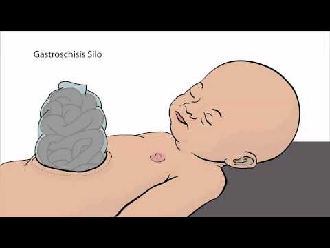
The surgical technique employed may vary depending on the size of the defect and the extent of intestinal herniation. In some cases, a silo may be created to gradually reduce the herniated intestine over a period of several days. Once the intestine has been reduced, the abdominal wall defect is repaired using a combination of sutures and mesh.
Intestinal hernia reduction is a critical component of surgical repair, as it helps to prevent further complications such as intestinal ischemia and necrosis. The surgeon must carefully assess the viability of the herniated intestine and resect any non-viable segments to ensure optimal outcomes.
In experienced hands, surgical repair of gastroschisis can be performed safely and effectively, with good outcomes and minimal complications. However, the procedure requires careful planning and execution to ensure optimal results.
Postoperative Care and Management
Following surgical repair of gastroschisis, postoperative care and management are critical to ensure optimal outcomes. The infant is typically admitted to the neonatal intensive care unit (NICU) for close monitoring and management.
Pain management is a priority, with analgesics administered as needed to minimize discomfort and promote healing. The infant is also closely monitored for signs of infection, such as fever, tachycardia, and elevated white blood cell count.
Nutritional support is also essential, with parenteral nutrition often initiated in the immediate postoperative period. As the infant recovers, enteral feeds are gradually introduced, with careful monitoring of feeding tolerance and stool output.
Serial abdominal X-rays are obtained to monitor for signs of intestinal obstruction or other complications. The infant is also closely monitored for signs of respiratory distress, with supplemental oxygen therapy administered as needed. Close collaboration between the surgical, neonatal, and nursing teams is essential to ensure seamless care and optimal outcomes.
Early recognition and management of postoperative complications are critical to preventing long-term sequelae and ensuring the best possible outcomes for infants with gastroschisis.
Long-term Outcomes and Follow-up Care
Infants with gastroschisis require long-term follow-up care to monitor for potential complications, such as intestinal obstruction, and to assess growth and development. Regular pediatric check-ups and abdominal ultrasounds are essential for optimal management.
Infant Health and Pediatric Care
Infants with gastroschisis require specialized pediatric care to manage their unique health needs. A multidisciplinary team of healthcare professionals, including pediatricians, surgeons, and nurses, should be involved in their care. Regular monitoring of infant health is crucial to identify potential complications, such as feeding intolerance, growth restriction, and intestinal obstruction.
Pediatric care for infants with gastroschisis should also focus on promoting optimal growth and development. This may involve nutritional support, physical therapy, and developmental assessments. Parents and caregivers should be educated on how to provide appropriate care and support for their infant, including managing feeding tubes, administering medications, and recognizing signs of complications.
Additionally, infants with gastroschisis may require ongoing surgical care to manage any long-term complications that may arise. This may include additional surgeries to repair intestinal hernias or to manage bowel obstruction. Close collaboration between pediatricians, surgeons, and other healthcare professionals is essential to ensure optimal outcomes for these infants.
Birth Defects and Developmental Delays
Children with gastroschisis are at increased risk of birth defects and developmental delays. Research suggests that up to 30% of children with gastroschisis may have associated birth defects, including cardiac, genitourinary, and musculoskeletal anomalies.
Developmental delays are also a concern, particularly in the areas of cognitive, motor, and language development. Studies have shown that children with gastroschisis may experience delays in reaching milestones, such as sitting, standing, and walking. Additionally, they may be at increased risk of learning disabilities and behavioral problems.
It is essential for healthcare providers to closely monitor the development of children with gastroschisis and provide early interventions to address any delays or deficits. This may include referrals to specialists, such as speech therapists, occupational therapists, and psychologists, to provide targeted support and therapy. By identifying and addressing these issues early, healthcare providers can help optimize outcomes and improve the quality of life for children with gastroschisis.
In conclusion, gastroschisis is a complex and multifaceted condition that requires a comprehensive and multidisciplinary approach to management. While significant advances have been made in the diagnosis and treatment of this condition, there remains a need for ongoing research and education to optimize outcomes and improve the quality of life for affected individuals.
Healthcare providers must work together to provide seamless care and support to families affected by gastroschisis, from prenatal diagnosis to long-term follow-up care. This includes ensuring access to specialized care, providing emotional support and counseling, and facilitating connections to community resources and support networks.
Ultimately, the goal of care for individuals with gastroschisis is to promote optimal health, development, and well-being. By working together and staying at the forefront of advances in medical knowledge and technology, we can make a meaningful difference in the lives of those affected by this condition and help them thrive.
This article provides a comprehensive overview of gastroschisis, including its causes, risk factors, and implications for maternal and fetal health. The discussion on genetic predisposition is particularly insightful.